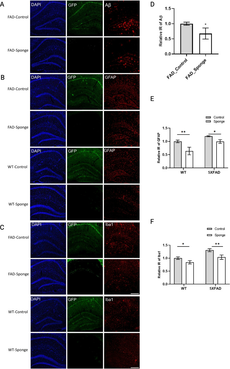
Fig. 3.
miR-29a sponge attenuates amyloid deposition and decreases activated astrocytes and microglia in the hippocampus of 5×FAD mice and WT mice. A–C Representative immunofluorescence images. D–F Quantification of Aβ, GFAP, and Iba1 immunoreactivity. Data were analyzed via two-way ANOVA (genotype by group) with Sidak’s post hoc tests. Bars indicate mean ± SD. N = 3 per group. Scale bars represent 100 μm; *p < 0.05, **p < 0.01, compared with the control group

