Fig. 3.
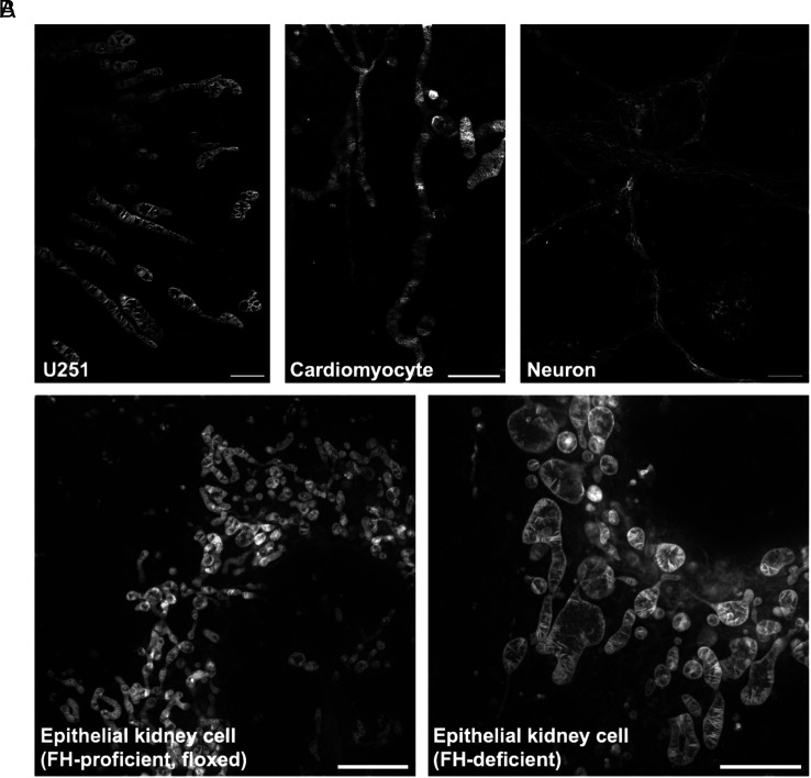
PKMO FX is a general mitochondrial probe for different cell lines. (A) PKMO FX-labeled mitochondria on different types of cells. From Left to Right: postfixation STED images of U251 cells, neonatal rat cardiomyocytes, and neuron cells. (Scale bars, 2 μm, 2 μm, 5 μm.) (B) STED images of FH-proficient (floxed) mouse epithelial kidney cells (Left) and FH-deficient cells (Right). In Fh1-KO cells, mitochondria show significantly swollen or swollen-elongated phenotypes with wider cristae spacing, uneven distribution of cristae, and the appearance of large hollow cavities compared to wild-type cells. (Scale bars, 5 µm.)

