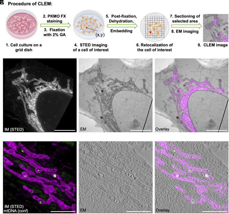
Fig. 6.
Multiplexed STED-CLEM using PKMO FX. (A) Experimental procedure of pre-embedding CLEM. (B) Left: STED image of a PKMO FX-labeled, 2% GA-fixed HeLa cell. Middle: TEM image of the same area. Right: Overlay of the FM and EM images. (Scale bars, 5 µm.) (C) Two-color CLEM experiment. Left: STED image of a PKMO FX-labeled, 2% GA-fixed HeLa cell (magenta). Background-subtracted confocal image of mtDNA (labeled with SYBR Gold, green). Middle: Single image extracted from an electron tomography stack. Right: Overlay of the two FM images with the EM image. (Scale bars, 3 µm.)

