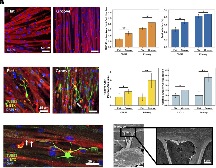
Fig. 2.
Effect of substrate topology on recreation of neuron-innervated muscle. (A) Immunofluorescence images of myotubes (MHC, red) and nucleus (DAPI, blue) of primary myoblasts differentiated on flat and microgrooved (Groove) substrates. (B) MHC-positive area divided by total cell number and (C) fusion index of C2C12-derived or primary myoblast-derived myotubes. (n = 5, *P < 0.05). (D) Immunofluorescence images of the neuron-innervated muscle made with primary myoblasts. [myotubes (MHC, red), neurons (TUBB3, green), AChRs (α-BTX, yellow), and nuclei (DAPI, blue)] (E) Innervation of neuron on primary myoblast-derived myotube on a grooved substrate. The white arrows indicate the innervation spot of the axonal terminal toward AChR clusters. (F) Relative AChR expression area on myotubes. (G) Relative colocalization area of axon and AChR on innervated muscles. (n ≥ 3, *P < 0.05, **P < 0.01). (H) Scanning electron microscopy (SEM) images of the innervated muscle.

