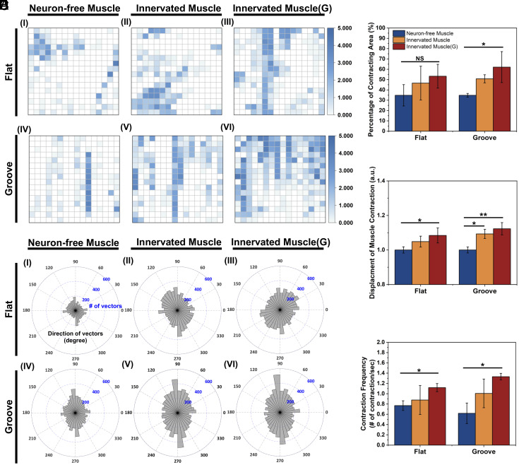
Fig. 3.
Analysis of muscle contraction of Neuron-free Muscle, Innervated Muscle, and Innervated Muscle(G), created on a flat and a microgrooved (Groove) substrate. (A) Visualization of the muscle contraction on the flat and the grooved substrate. Size of the view: 299.5 μm × 224.6 μm. The z value represented the number of contractions in each block per second. (B) Percentage of contracting myotubes. (C) Relative contraction displacement of myotubes. The “y” value was normalized to the value of Neuron-free Muscle on the flat substrate. (D) Direction of muscle contraction 90 and 270 degrees were aligned with the grooved pattern direction. (E) Average contraction frequency of the muscles (n= 3, *P < 0.05, **P < 0.01).

