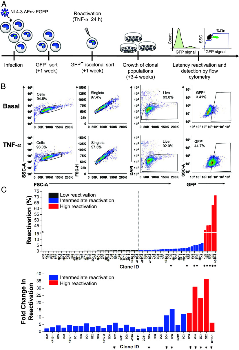
Fig. 1.
Generation of a monocyte model of HIV-1 latency. (A) THP-1 monocytes were infected with HIV-1 NL4-3 ΔEnv EGFP. After 1 wk, GFP- cells were sorted by FACS and stimulated with TNF-α for 24 h the following week. GFP+ cells were individually sorted and allowed to relax back into a latent state to generate clonal TLat cell lines. Clonal cell lines were analyzed for GFP expression after TNF-α stimulation. (B) Representative flow cytometry gating and analysis for basal (Top), and TNF-α (Bottom) stimulated conditions. Gating was set based on the uninfected THP-1 population (SI Appendix, Fig. S1A). (C) HIV latency reactivation percentage of the TLat clonal library after TNF-α stimulation for 24 h (Top). Fold change in reactivation of the intermediate and high reactivation clones (Bottom). Clones denoted with asterisks were selected for further analysis.

