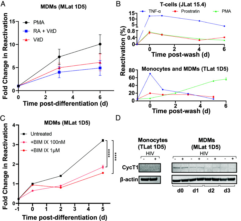
Fig. 3.
Mechanisms of HIV reactivation during macrophage differentiation. (A) Reactivation postdifferentiation was evaluated in MLats (1D5 clone) generated by different methods (PMA, VitD, and RA+VitD). Data are represented as mean of five replicates from two independent experiments ± SEM. (B) Three LRAs (PKC agonists) TNF-α, Prostratin, and PMA, were added to latently infected T-cells (Top) and monocytes (Bottom) for 24 h and removed (t = 0) to evaluate HIV reactivation over time. PMA causes MDM differentiation in monocytes. Data represents the mean of at least three independent replicates ± SEM. (C) Latently infected monocytes were pre-treated with the nonselective PKC inhibitor BIM IX for 30 min prior to differentiation into macrophages and for the duration of the experiment. HIV reactivation was quantified at different time-points following differentiation. Statistical significance was determined by performing a two-way ANOVA comparison with Dunnett correction (****P < 0.0001). Data represents the mean of three independent replicates ± SEM. (D) Western blot analysis of cyclin T1 (CycT1) and B-actin control in THP-1 (−HIV) and TLat 1D5 (+HIV) monocytes and MDMs.

