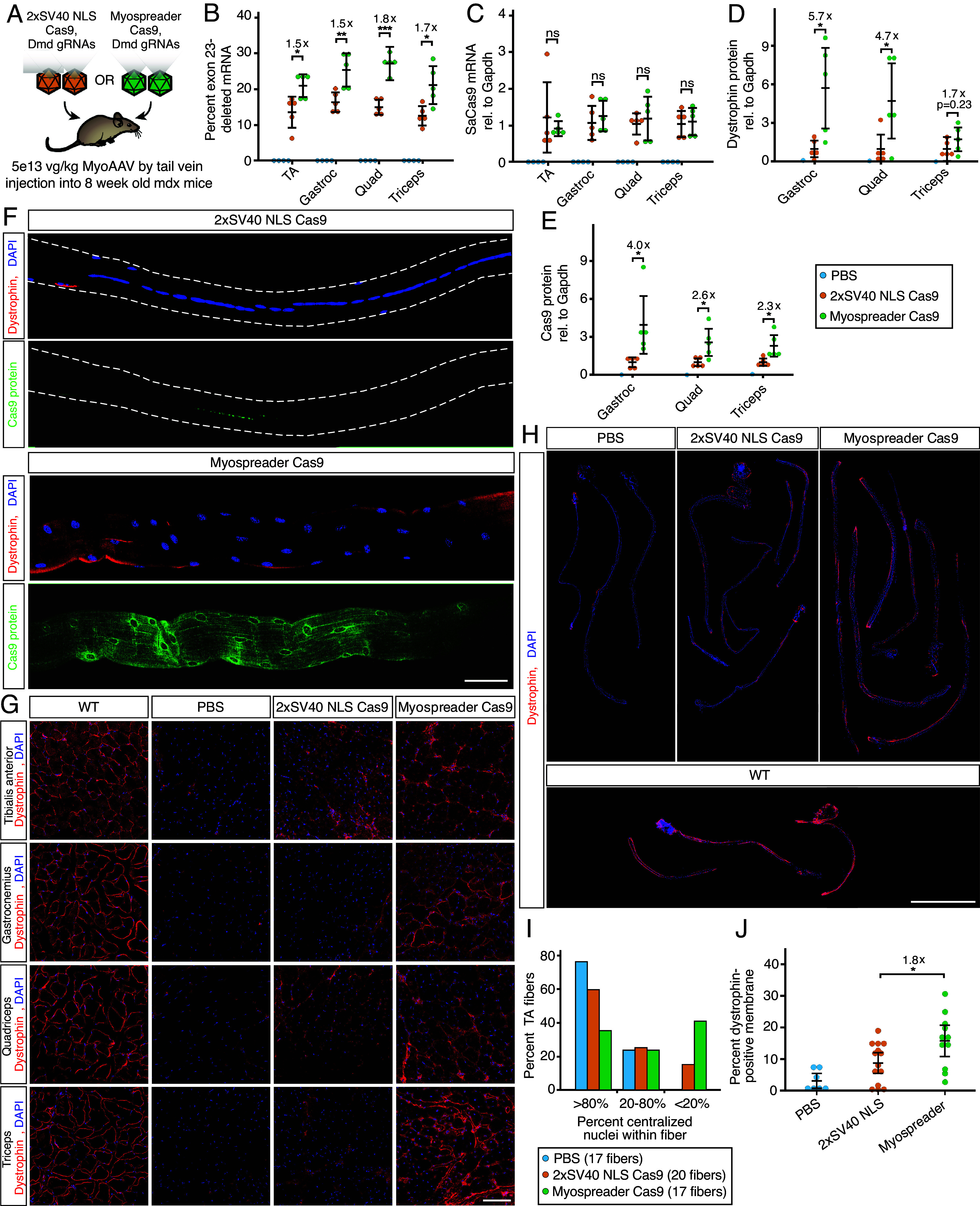
Fig. 4.

Myospreader improves Cas9 editing efficiency in a Mouse model of Duchenne muscular dystrophy. (A) Schematic of the experiment to measure the efficacy of AAVs containing 2xSV40 NLS Cas9 or Myospreader Cas9, systemically delivered to 4-wk-old mdx mice at a dose of 5E+13 vg/kg. (B) RT-qPCR quantification of exon-23 deleted Dmd mRNA in different muscles of treated mdx mice. Significance by Student’s t test. (C) RT-qPCR quantification of Cas9 mRNA in different muscles of treated mdx mice. Plotted relative to the mean of the 2xSV40 NLS Cas9 group. Significance by Student’s t test. (D) Quantification of dystrophin protein as determined by western blot/densitometry, normalized to Gapdh. Plotted relative to the mean of the 2xSV40 NLS Cas9 group. Significance by Student’s t test. (E) Quantification of Cas9 protein as determined by western blot/densitometry, normalized to Gapdh. Plotted relative to the mean of the 2xSV40 NLS Cas9 group. Significance by Student’s t test. (F) IF to detect dystrophin (red) and Cas9 (green) protein in representative TA myofibers isolated from treated mdx mice. (Scale bar: 50 µm.) (G) Representative images of Dystrophin protein (red) and DAPI (blue) in muscle cryosections of treated mdx mice by IF. (Scale bar: 100 µm.) (H) IF to detect dystrophin protein (red) and DAPI (blue) in whole TA myofibers isolated from treated mdx mice or untreated 8-wk-old WT mice. (Scale bar: 1 mm.) (I) Quantification of centralized nuclei in TA myofibers isolated from treated mdx mice. (J) Quantification of dystrophin protein at the periphery of TA myofibers of treated mdx mice or 8-wk-old WT mice. Significance determined Student’s t test (ns = not significant; *P < 0.05; **P < 0.01; ***P < 0.001) (error bars = 95% CI).
