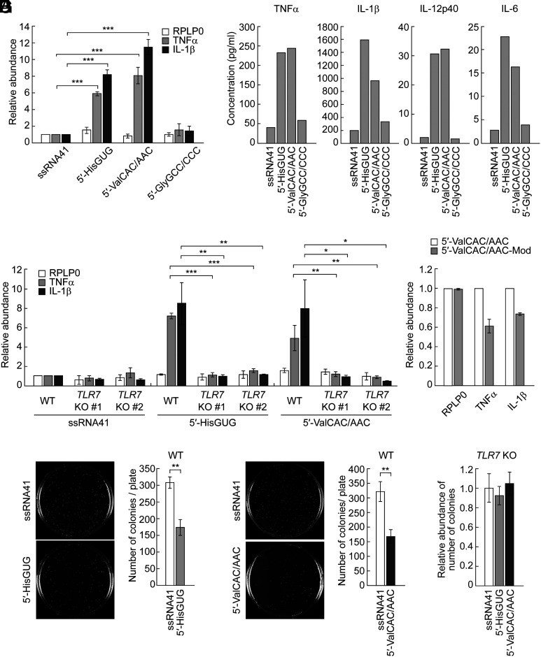
Fig. 1.
The 5′-tRNAValCAC/AAC half activates TLR7. (A) Using DOTAP, 5′-tRNA halves and ssRNA41 (negative control) were transfected into HMDMs. Total RNAs from the cells were subjected to RT-qPCR for the indicated mRNAs. The quantified mRNA levels were normalized to the levels of GAPDH mRNA. For all graphs in the present study, error bars indicate mean ± SD of triplicate measurements (*P < 0.05, **P < 0.01, and ***P < 0.001; two-tailed t test). (B) After RNA transfection into HMDMs using DOTAP, the culture medium was subjected to measurement of concentration of the indicated cytokines. Means of two independent experiments are shown. The experimental results from three replicates for both the 5′-tRNAHisGUG half and the 5′-tRNAValCAC/AAC half are presented in Fig. 3B, while those for the 5′-tRNAGlyGCC/CCC half are shown in SI Appendix, Fig. S1. (C and D) The experiments in (A) were performed by using two different TLR7 KO THP-1 cell clones (C) or the 5′-tRNAValCAC/AAC half with modifications (D). (E and F) HMDMs transfected with the 5′-tRNAHisGUG half (F) or the 5′-tRNAValCAC/AAC half (G) were subjected to bacterial infection and invasion assay. Transfection of ssRNA41 was used as a negative control. Representative pictures of the plates with E. coli colonies and bar graphs of the counted numbers of colonies are shown. (G) The experiments in (F) and (G) were performed by using TLR7 KO THP-1 cell #1.

