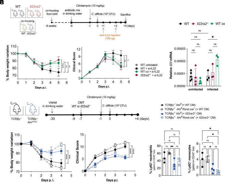
Fig. 3.
Production of IL-22 during C. difficile infection is not required to protect Il22ra2–/– mice. (A) Schematic representation of C. difficile infection and IL-22 blockade by i.p. injection of anti–IL-22 neutralizing antibody on days 1 and 3 p.i. (B) Body weights (Left) and clinical scores (Right) of WT and Il22ra2–/– mice. (C) Relative expression of Il22 mRNA in total proximal colon of naïve WT and Il22ra2–/– mice, whether cohoused or not since birth, normalized by Gapdh. (D) Schematic representation of CMT from Il22ra2–/– into TCRβδ–/– Ahrfl/fl (T cell-deficient) and TCRβδ–/– Ahrfl/fl RORγt-Cre (T cell- and ILC3-deficient) mice. (E) Body weights (Left) and clinical scores (Right) of TCRβδ–/– Ahrfl/fl and TCRβδ–/– Ahrfl/fl RORγt-Cre mice that received WT or Il22ra2–/– CM before C. difficile infection. (F) FACS quantification of CD11b+ Ly6G+ neutrophils (Left) and CD11b+ Ly6C+ inflammatory monocytes (Right) in the colonic lamina propria of mice in (E) on day 4 p.i. (A and B) n = 5, (C) n = 6, and (D and E) n = 3 to 4 mice per group. (B and E) Two independent experiments are combined. Error bars represent mean ± SEM. Normality was assessed by D’Agostino–Pearson test; statistical analysis was performed using One-way ANOVA with post hoc Tukey test. *P < 0.05; **P < 0.01; ***P < 0.001; ****P < 0.0001; ns = not significant.

