Fig. 1.
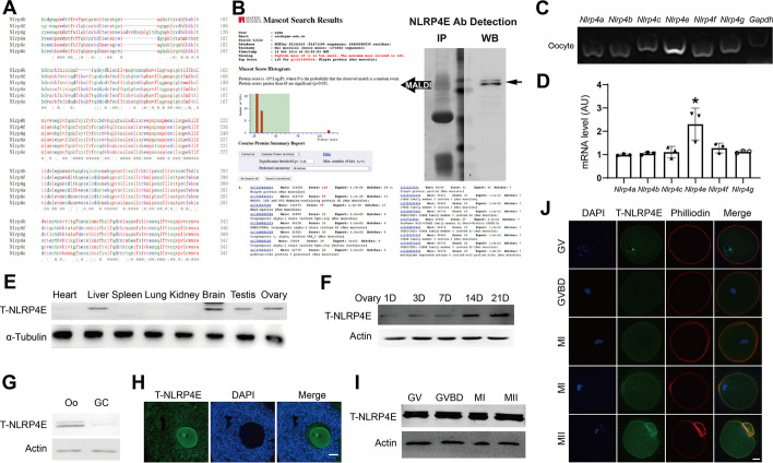
NLRP4E is an oocyte-predominant protein. A The AA sequence alignment between NLRP4 family members, including the 75–323 AA region in NLRP4E for construction of the polyclonal antibody used in this study. B Verification of the specificity of NLRP4E polyclonal antibody. We used NLRP4E antibody to perform immunoprecipitation followed by SDS-PAGE and silver staining. The gel band (left) corresponding to the NLRP4E blot band (right) was cut and sent for MALDI. The results show that the identified protein with maximum intensity was NLRP4E. C, D RT-PCR shows that NLRP4E mRNA was the richest among NLRP4 family mRNAs. E Western blot showing that NLRP4E was predominantly expressed in the brain, liver, testis, and ovary. F Western blot showing that the NLRP4E level gradually increased in mouse ovaries from post-natal day (PND) 1 to 21 and reached a peak on PND 21. G, H Western blots (G) and immunofluorescence (H) show that NLRP4E was more predominant in oocytes than in GCs (granular cells). I Western blots show that the NLRP4E level remained constant during oocyte meiosis. J Immunofluorescence shows that NLRP4E accumulates at the cell membrane during oocyte meiosis. DAPI is shown in blue, NLRP4E in green, and actin filaments in red. GV, germinal vesicle; GVBD, germinal vesicle breakdown; MI, metaphase I; MII, metaphase II. Scale bar, 50 μm in H, 20 μm in J. * indicates p < 0.05

