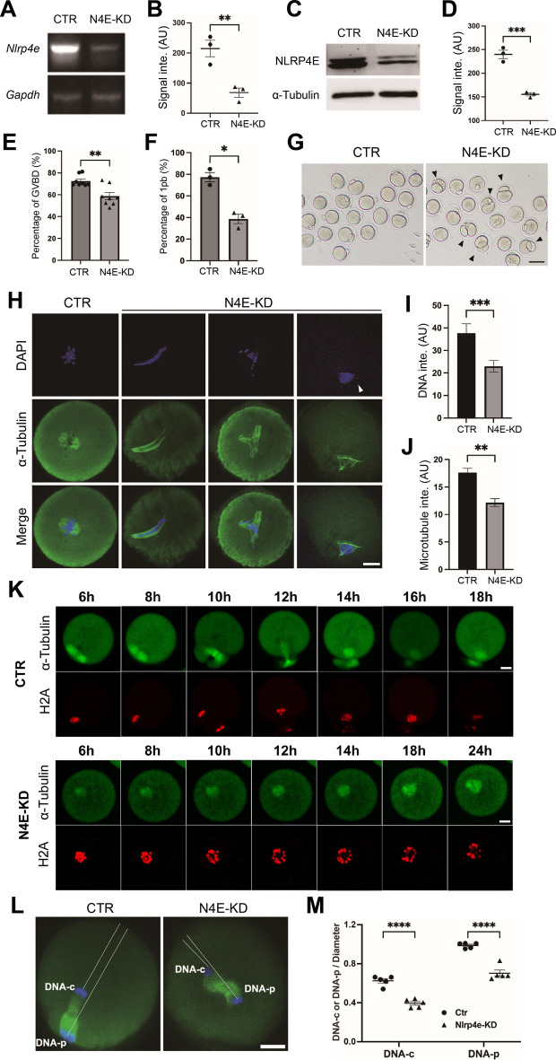
Fig. 2.
NLRP4E knockdown disrupts oocyte meiosis. A, B RT-PCR and quantification showing that the NLRP4E mRNA level was significantly reduced by siRNA knockdown. C, D Western blotting shows that the NLRP4E level was significantly reduced by siRNA knockdown. E–G Quantification shows that NLRP4E knockdown significantly reduced the percentages of GVBD (E) and 1pb (the first polar body, F and G). H–J Immunofluorescence analysis shows that NLRP4E knockdown caused significant disruption of chromosomes and spindle microtubules. DNA is shown in blue and microtubules are in green. K Slices from live imaging show that in some oocytes with severe NLRP4E knockdown, the chromosomes failed to segregate at the end of meiosis. EGFP-tubulin mRNA and TagRFP-histone were injected into oocytes to label microtubules and chromosomes. L, M Immunofluorescence analysis shows that in oocytes with medium NLRP4E knockdown, the chromosomes could segregate at the end of meiosis, but spindle translocation toward the cortex was significantly blocked. Scale bar, 80 μm in G and 20 μm in other panels. * indicates p < 0.05; ** indicates p < 0.01; *** indicates p < 0.001; **** indicates p < 0.0001

