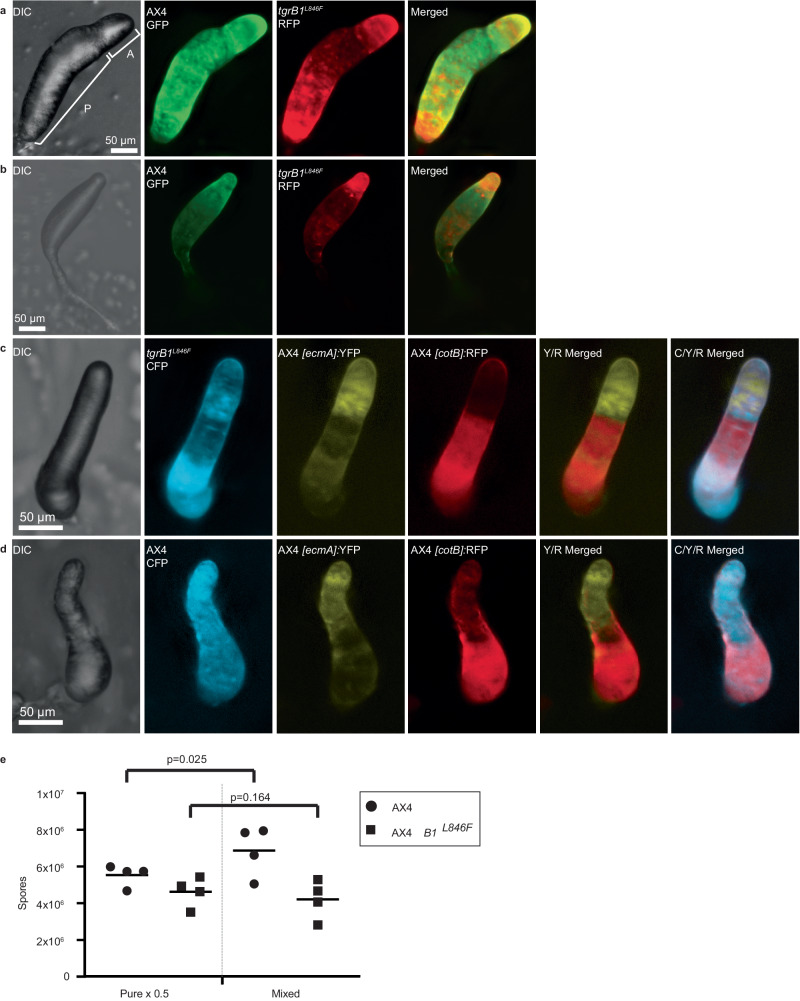
Fig. 1. Activated tgrB1 confers altruistic behavior.
We used two strains that express constitutive fluorescent markers, the wild-type AX4-GFP (green) and the activated tgrB1 mutant AX4 tgrB1L846F-RFP (red). We grew the cells separately, mixed equal proportions, and co-developed them. We imaged the structures at the finger (a) and culminant stage (b) with DIC and with green and red fluorescence, and generated a merged image of the red and green channels as indicated. The brackets in panel a show the anterior region (A) that contains mainly prestalk cells and the posterior region (P) that contains mainly prespore cells. c We also co-developed constitutively labeled mutant tgrB1L846F-CFP cells (cyan) with wild-type AX4 cells carrying the prestalk reporter [ecmA]:YFP (yellow) and the prespore reporter [cotB]:RFP (red). We imaged the structures with DIC and with cyan, yellow, and red fluorescence, and generated merged images of the yellow and red (Y/R) as well as all three channels (C/Y/R) as indicated. d As a control, we co-developed constitutively labeled wild-type AX4-CFP cells (cyan) with wild-type AX4 cells carrying the same prestalk and prespore reporters and imaged them as above. e We grew wild-type AX4-GFP and mutant AX4 tgrB1L846F-RFP cells separately, developed 7×10⁶ cells either in pure populations or mixed at equal proportions as indicated, and counted spores. The spore counts are shown as four independent replicates (symbols) and their averages (horizontal lines). The pure population counts were multiplied by 0.5 to scale them with the mixed population. Brackets and p-values (T-test, one-sided, n = 4) compare the spore counts of each strain in the two conditions. Camera settings are included in Supplementary Data 1. Source data are provided as a Source Data file.

