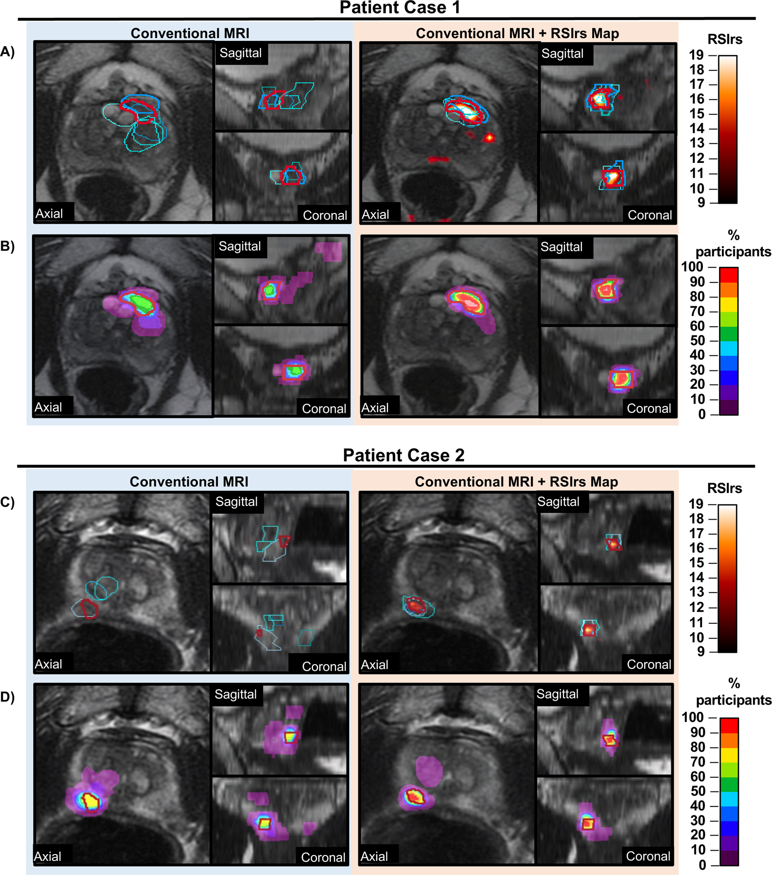
Figure 1: RSIrs maps improve the accuracy of participant target volumes.

Example target volumes from two patient cases (Case 1: A/B and Case 2: C/D) where participants were provided Conventional MRI alone (T2-weighted, DWI, ADC) (left) images or conventional MRI with RSIrs map (orange heat map) (right). Expert volumes are shown in red in all panes. Select participant volumes are highlighted in shades of blue in (A and C). All participant volumes for each case are displayed in (B and D) as a rainbow heatmap, where the color represents the percentage of participants who included that voxel in their target volume.
