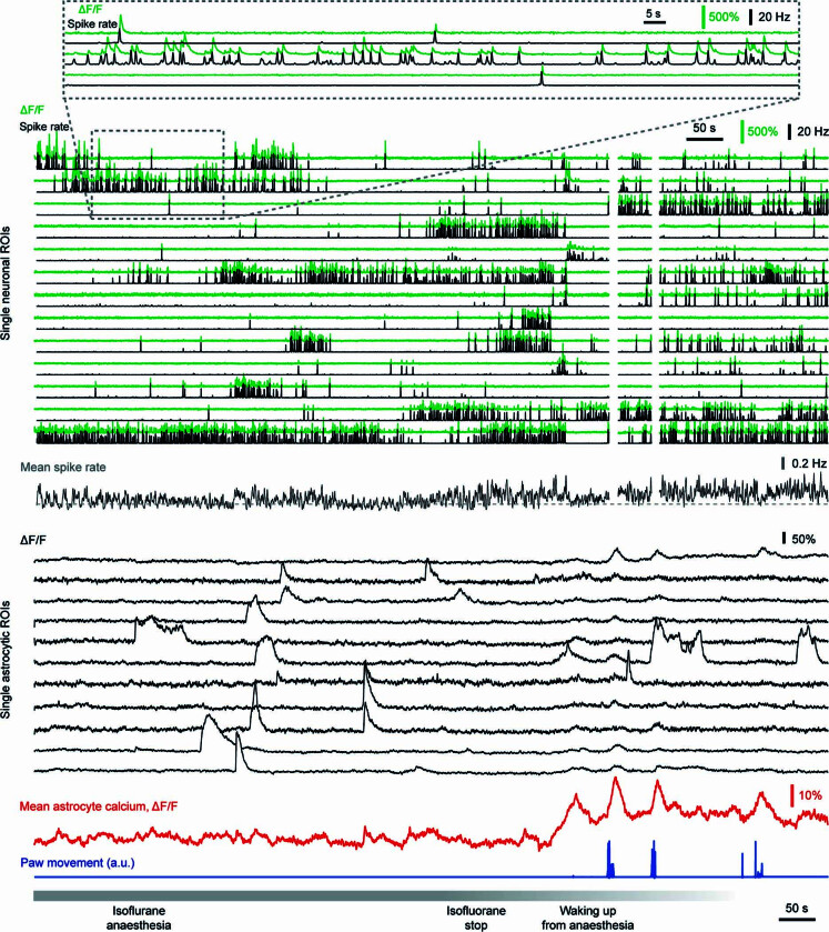
Extended Data Fig. 10. Calcium imaging of astrocytes and pyramidal neurons in CA1 during isoflurane anesthesia.
Examples of extracted neuronal ΔF/F traces (green) and associated deconvolved spike rates (black) are shown (see also zoom-in at the top). White blanked time points were discarded due to excessive movement of the brain during waking up. Example astrocytic ΔF/F traces (black) are shown below, highlighting uncoordinated local but no global events before waking up. Waking up (bottom; 1.5% in the beginning, set to 0% at ‘isoflurane stop’) is reflected by small and large paw movements (blue) and resulted in global astrocytic calcium signals (mean astrocytic calcium, red).

