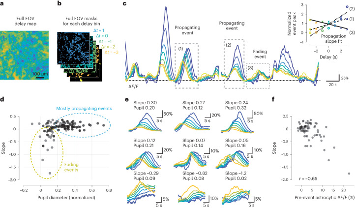
Fig. 6. Centripetal propagation of activity in astrocytes is conditional on arousal state and cell-intrinsic calcium signaling history.
a,b, A smoothed delay map of the entire FOV (a) is binned according to the delays of each pixel (b) (Δt = 1 corresponds to the 0.5 … 1.5 s interval). c, The delay bin masks (b) are used to extract global astrocytic traces from pixels with a specific delay from the denoised imaging data (color coding). Due to centripetal propagation, yellow traces represent gliapil and blue traces astrocytic somata. Some calcium events propagate to somata (events (1) and (2)), while others do not (event (3)), quantified by a positive or negative propagation slope, respectively (inset). Therefore, centripetally propagating and nonpropagating events can be determined by the slope of peak activity with respect to delay. d, Propagation slope of events plotted against the normalized pupil diameter as a proxy for arousal state. Each data point corresponds to a single event. e, Example events are plotted in the order of sorted slope values. See Extended Data Fig. 7 for additional example events. f, Pre-event astrocytic ΔF/F, averaged across the 20 s before the event, is negatively correlated with the slope value. Only data points with normalized pupil diameter < 0.2 from d were included for f.

