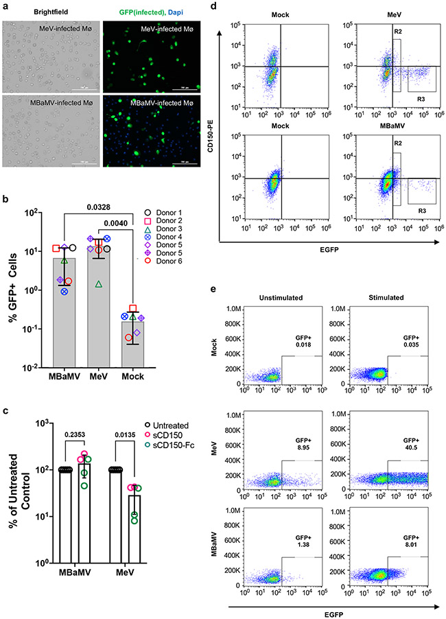
Figure 4. MBaMV infects human monocyte-derived macrophages (MDM) in a CD150-independent manner.
a-b, MDMs were infected with MV323-EGFP or MBaMV (1x10⁵ IU/sample) and were either (a) fixed by 2% PFA at 24 hpi, DAPI-stained and imaged (scale bar is 200 μm), or (b) quantified by flow cytometry. The percent of CD68+GFP+ MDMs from 6 donors are shown. Open and crossed symbols indicate experiments using lot 1 and lot 2 viruses, respectively. Adjusted p values are from one way ANOVA with Dunnett’s multiple comparisons test. c, Soluble human CD150 (sCD150) or a CD150 Avi-tag inhibited MeV but not MBaMV infection of macrophages. GFP+ events in untreated controls were set to 100%, and entry under sCD150/ CD150 Avi-tag were normalized to untreated controls. Adjusted p values are from two-way ANOVA with Šídák’s multiple comparisons test. In (b) and (c), data shown are mean +/− S.D. from multiple experiments (n=7 and n=5, respectively) with individual values also shown. (d) Exemplar FACS plots from the summary data shown in (b) for CD150 staining. e, ConA/IL-2 stimulated PBMCs were infected with MeV or MBaMV (MOI of 0.1) and analyzed for GFP expression by flow cytometry at 24 hpi.

