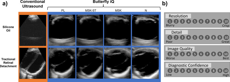
Fig 4. Example images and Likert scale featured in the survey.
a) Select images of pathologies featured in the survey, including intraocular silicone oil and tractional retinal detachment. COU images are seen in the orange outline, whereas Butterfly iQ images with the PL, MSK-ST, MSK, and N presets are displayed with the blue outline. b) Likert scale used by graders to evaluate images for resolution, detail, image quality, and diagnostic confidence.

