
Figure 1.
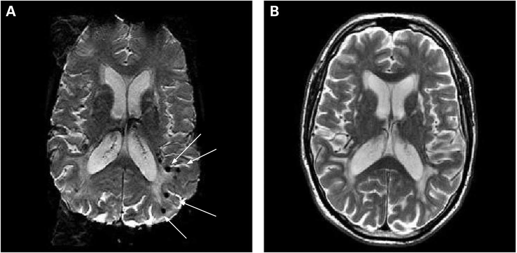
Cerebral microbleeds (CMBs), as seen on T2* weighted gradient echo type echo planar (GRE-EPI) MRI. To differentiate from areas of signal loss based on vascular flow voids, CMBs are scored if there is a focal area of signal loss on T2* weighted GRE-EPI images (A, arrows) that are invisible or smaller on T2 weighted fast spin echo images (B).