
Fig. 1 |. Mitochondrial volume and function are compromised in the absence of dFMRP.
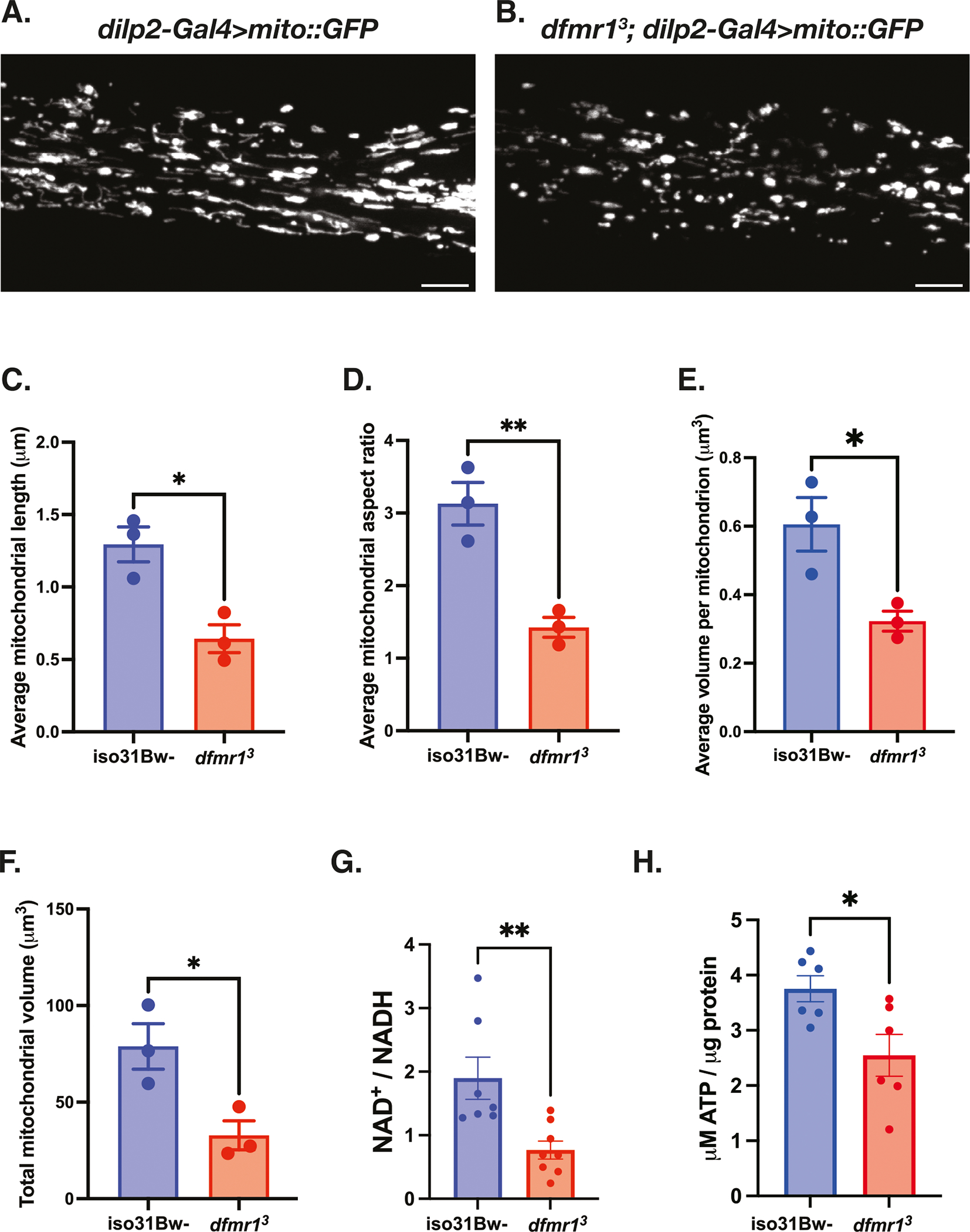
A–F Mitochondria in the insulin producing cells (IPCs) of the brain were labeled by expressing a genetically encoded UAS-mitoGFP construct in conjunction with the dilp2-Gal4 driver. Representative maximum-intensity projections of GFP-labeled mitochondria in the IPC processes of (A) iso31Bw- wild type and (B) dfmr1 mutant flies. Scale bars: 5 μm. Images are oriented with the dorsal side on the left and the ventral side on the right. Quantification of (C) average mitochondrial length, (D) average mitochondrial aspect ratio, (E) average volume per mitochondrion, and (F) average total mitochondrial volume per brain. Sample number (N) per genotype = 3 brains. Unpaired t-tests indicated that the average length, aspect ratio, volume per mitochondrion, and total mitochondrial volume per brain were all significantly reduced in dfmr1 mutants compared to iso31Bw- wild type controls. Values represent mean ± SEM. *p ≤ 0.05, **p ≤ 0.01. G Quantification of the NAD+/NADH ratio. Each sample contained 10 fly bodies. Sample number (N) per genotype: iso31Bw- = 7, dfmr1 = 8. An unpaired t-test indicated that the NAD+/NADH ratio was significantly diminished in dfmr1 mutants compared to iso31Bw- wild type controls (p = 0.0059). Values represent mean ± SEM. H Quantification of ATP levels relative to protein content. Each sample contained 5 fly bodies. Sample number (N) per genotype = 6. An unpaired t-test showed that ATP levels were significantly decreased in dfmr1 mutants compared to iso31Bw- wild type controls (p = 0.0224). Values represent mean ± SEM.