
Figure 5:
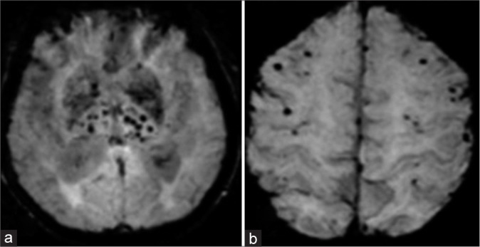
(a) Susceptibility-weighted images demonstrating cerebral microbleeds in the deep brain regions including basal ganglia, thalamus, and brainstem (b) in a case of hypertensive arteriopathy; peripheral lobar distribution in a patient with cerebral amyloid angiopathy.