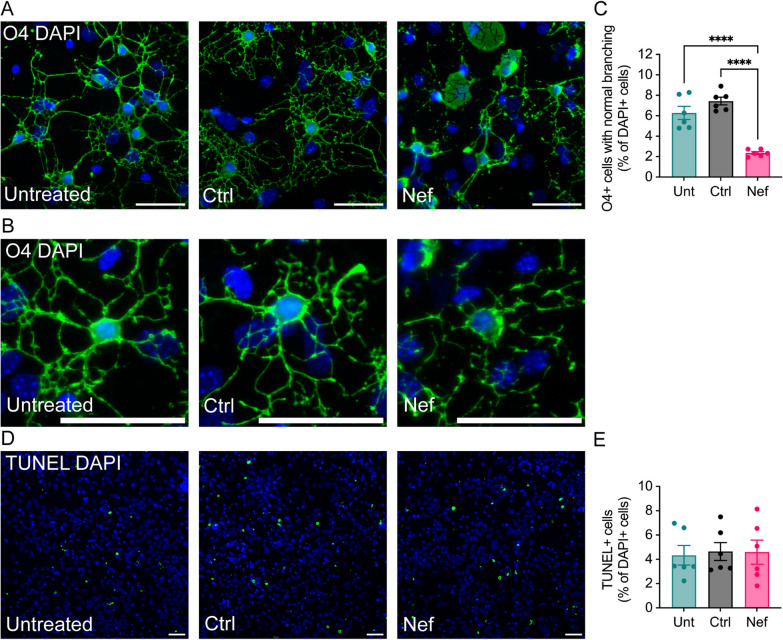
Fig. 5.
Effects of Nef EVs on oligodendrocytes in primary brain cultures. Mouse primary brain cells were collected at postnatal day 3 and grown for 5 days in media to promote oligodendrocyte growth. Subsequently, the cells were treated with either Ctrl or Nef EVs for 48 h. A, B Untreated (Unt) cultures served as negative controls. Representative fluorescence images illustrate oligodendrocytes (O4, green) and nuclei (DAPI, blue). Higher magnification images displaying branching oligodendrocyte morphology are presented in (B). C Quantification of O4 + oligodendrocytes with clear immunostaining and normal branching as a proportion of total DAPI + cells. D Representative fluorescence images of the TUNEL assay. E Quantification of the TUNEL assay. Scale bar is 50 µm. N = 6, ****p < 0.0001

