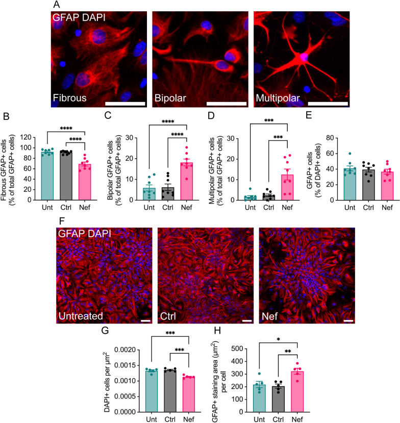
Fig. 6.
Effects of Nef EVs on astrocyte morphology in vitro. Mouse primary brain cells were collected at postnatal day 3. After 5 days of growth in media to promote oligodendrocyte development, cells were treated with either Ctrl or Nef EVs for 48 h, with untreated (Unt) cultures served as negative controls. A. Representative fluorescence images depict the three primary types of astrocyte morphology observed, including (i) fibrous, (ii) bipolar, and (iii) multipolar. GFAP (red) and DAPI (blue) label astrocytes and nuclei, respectively. B–D Quantification of fibrous, bipolar, and multipolar GFAP + cells as a proportion of the total GFAP + cell population. N = 5 images/group. E Quantification of GFAP + cells as a proportion of DAPI + cells. F A2B5 + cells were purified via MACS from dissociated mouse brains, cultured for 3 days in media to promote astrocyte differentiation, and then treated with either Ctrl or Nef EVs for 48 h. GFAP (red) and DAPI (blue) label astrocytes and nuclei, respectively. Scale bar is 50 µm. G Quantification of DAPI + cells. H Quantification of GFAP + cells. N = 5, *p < 0.05; **p < 0.01; ***p < 0.001; ****p < 0.0001

