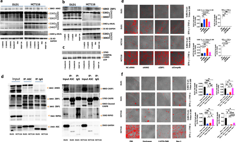
Fig. 8. Hyperactivation of PANoptosis effective molecules by AIM2-ZBP1-RIPK1-RIPK3-ASC-CASP8-CASP1 signal pathway.
Immunoblot analysis of AIM2, ZBP1, GSDMD, GSDME, p-MLKL in DLDL1 or HCT116 treated with siAIM2s, siZBP1s or siChmp4b (a–c). Immunoblot analysis of AIM2, ZBP1, RIPK1, ASC, CASP1, CASP8, RIPK3 in DLDL1 or HCT116 immunoprecipitated with IgG control antibodies or anti-ASC antibodies (d). GAPDH was used as the internal control. DLD1 and HCT116 were treated by 0.001% v/v Triton X-100 for 7 h or by IFN-γ + TNF-α for 24 h, after treatment respectively with NC-siRNA, siAIM2, siZBP1 or siChmp4b for 48 h. DLD1 and HCT116 were treated with 0.001% v/v Triton X-100 for 12 h after treatment with Emricasan alone, Z-IETD-FMK alone or Nec-1 alone for 24 h (e); or DLD1 and HCT116 were co-treated with IFN-γ + TNF-α, IFN-γ + TNF-α + Emricasan, IFN-γ + TNF-α + Z-IETD-FMK or IFN-γ + TNF-α + Nec-1 for 36 h (f). All cells were stained by PI, and time-lapse confocal images of PI + DIC. Two-tailed unpaired Student’s t-test was used to determine significance (*P < 0.05; **P < 0.01; ***P < 0.001; ****P < 0.0001; NS, not significant). Scale bars, 50 um. Data are representative of at least three independent experiments.

