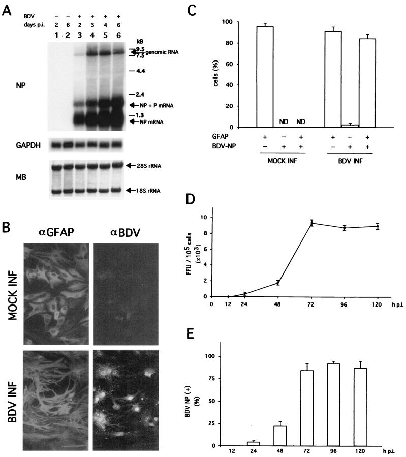
FIG. 1.
(A) Synthesis of BDV RNA in primary FeAst. Primary cat astrocytes were infected with BDV-He80 at an MOI of 0.1 FFU/cell. At the indicated times, total RNA from mock- and BDV-infected cells was analyzed by Northern blot hybridization. BDV genomic and NP mRNAs were detected with a 32P-labeled NP DNA probe. As a control, the same blot was also hybridized with a probe for the housekeeping cellular mRNA GAPDH. MB staining of the membrane after transfer and prior hybridization was used to verify that similar amounts of total RNA were loaded in all cases. The positions of the 28S and 18S rRNAs are indicated. (B) Detection of BDV NP antigen and GFAP expression in primary FeAst. BDV-infected cells (INF) and mock-infected controls were fixed at 96 h p.i. and analyzed by indirect immunofluorescence. Cells were simultaneously labeled with a rabbit antiserum to GFAP (αGFAP) and a rat serum to BDV (αBDV). (C) The percentage of cells expressing one or both antigens was determined by counting cells (total of 250) from five or six different fields. Shown are the average and standard deviation from two independent experiments. ND, not determined. (D) Kinetics of BDV multiplication in primary FeAst. Cells were infected with BDV-He80 at an MOI of 0.1 FFU/cell. At the indicated times p.i., BDV infectivity in supernatant and in WCE was determined. Only cell-associated infectivity was detected. Values correspond to the average and standard deviation of two independent experiments. (E) Number of BDV-infected cells at different times p.i. FeAst were infected at an MOI of 0.1 FFU/cell, and at the indicated times after infection, cells were fixed and examined for expression of BDV NP antigen by IIF. Values correspond to the average and standard deviation of two independent experiments.

