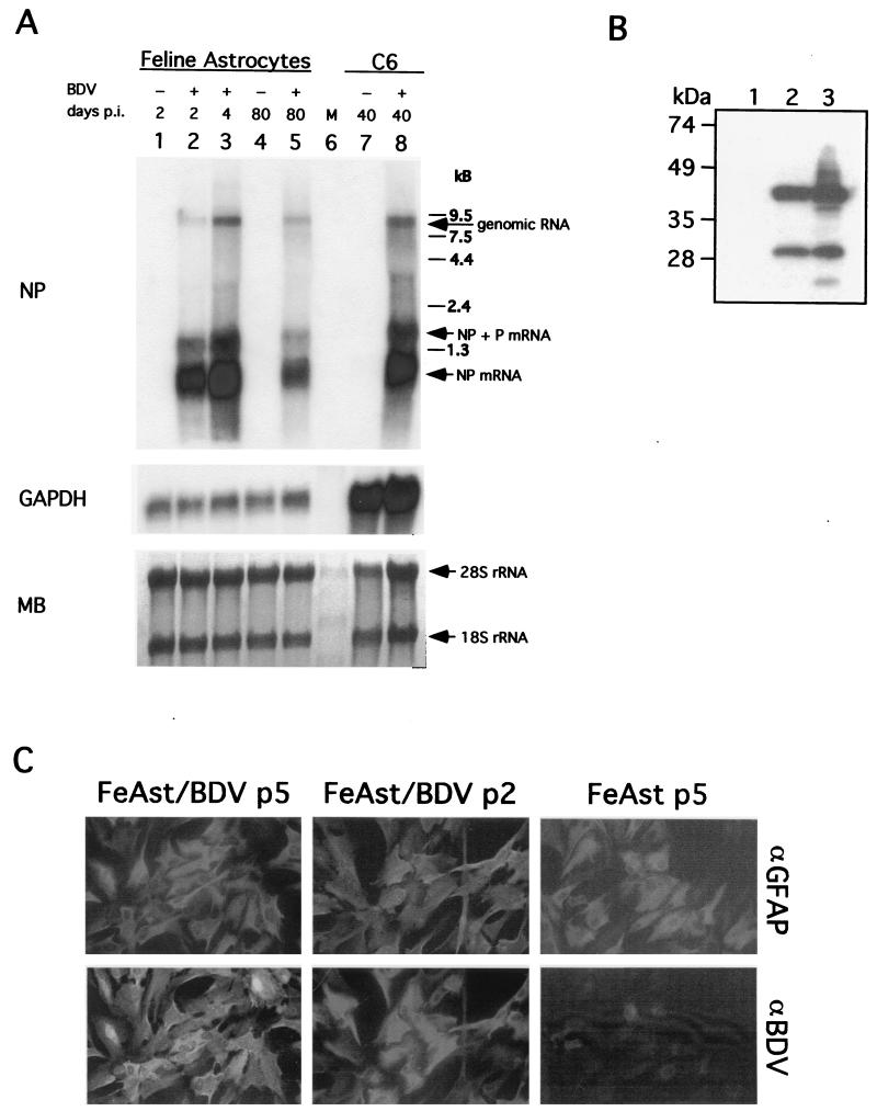
FIG. 2.
BDV persists in primary FeAst. (A) Detection of BDV RNA both genome and mRNA in FeAst at 80 days p.i. (passage 4). RNA was extracted from BDV-infected FeAst (lanes 2, 3, and 5) at 2, 4, and 80 days p.i. and from mock-infected control FeAst (lanes 1 and 4) at 2 and 80 days. RNA (5 μg each) was analyzed by Northern blot hybridization with probes for BDV NP and rat GAPDH. As a comparison, RNA was also extracted from C6 cells persistently infected with BDV. Differences in the hybridization signal with the GAPDH probe reflect nucleotide differences between cat and rat species in the GAPDH gene. MB staining of the membrane is shown. The positions of the 28S and 18S rRNAs are indicated. (B) Detection of BDV NP and P. WCE from FeAst-p5 (lane 1), FeAst/BDV-p5 (lane 2), and C6BV-p10 (lane 3) were analyzed by Western blotting as described in Materials and Methods. (C) Detection by IIF of BDV NP antigen in FeAst at 40 and 100 days p.i. BDV-infected and mock-infected control FeAst were fixed at days 40 and 100 p.i. and analyzed by IIF with double labeling with antibodies to GFAP (αGFAP) and BDV NP (αBDV).

