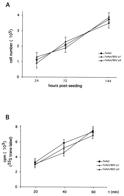
FIG. 3.
Cell growth and synthesis of proteins are not impaired in FeAst persistently infected with BDV. (A) Cell growth. Mock-infected (p4) and BDV-infected FeAst p1 and p4 were seeded (105 cells/dish) into 35-mm-diameter polylysine-treated tissue culture dishes. At the indicated postseeding times, the numbers of viable cells were determined by using trypan blue staining. For each sample and time point, cell numbers were determined in triplicate. Shown are average values and standard deviations of two independent experiments. (B) Rate of protein synthesis. Cells were labeled with 35S-Trans label for the amount of time indicated. Incorporation of 35S label into proteins was determined by trichloroacetic acid precipitation and Cerenkov counting with a scintillation counter. Each time point was determined in triplicate. Values correspond to the average and standard deviation.

