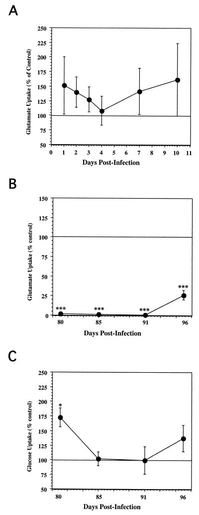
FIG. 5.
(A) Effects of BDV acute infection on FeAst glutamate uptake. At the indicated time points after infection with BDV, the 3H-Glu uptake assay was performed, as described in Materials and Methods. Results are expressed as the ratio of 3H-Glu uptake of infected cultures over that of control cultures. (B) Effects of BDV persistent infection on FeAst glutamate uptake. At the indicated time points after infection with BDV, the 3H-Glu uptake assay was performed, as described in Materials and Methods. Results are expressed as the ratio of 3H-Glu uptake of infected cultures over the value of the control cultures ± standard error of the mean. Results were analyzed by analysis of variance. ∗, P < 0.05; ∗∗, P < 0.01; ∗∗∗, P < 0.001. (C) Effects of BDV persistent infection on astrocyte glucose uptake. At the indicated time points with BDV, the ability of the feline astrocyte cultures to remove 3H-2DG from the culture supernatant was determined, as described in Materials and Methods. Results are expressed as the ratio of [3H]glucose uptake of infected cultures over the value of the control cultures.

