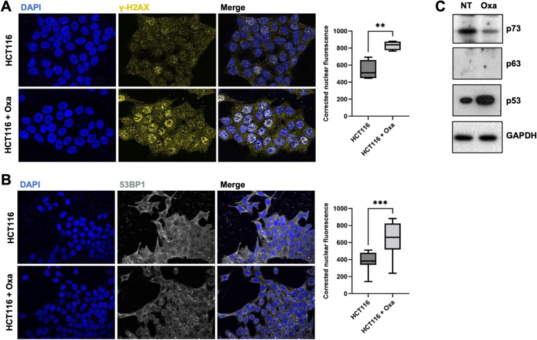
Fig. 1.
Oxaliplatin treatment induces DNA damage. A Immunofluorescence experiments performed on HCT116 cells treated, or not, with oxaliplatin (0.5 µM) monitoring γ-H2AX (yellow). DAPI (blue) was used to stain DNA and the nuclei. Quantification of the staining is shown on the right side and is represented as the corrected nuclear fluorescence. Data are represented as the mean ± SEM. (n = 3). Unpaired two-tailed Student’s t-test; ** p < 0.01. B Immunofluorescence experiments performed on HCT116 cells treated, or not, with oxaliplatin (0.5 µM) monitoring 53BP1 (grey). DAPI (blue) was used to stain DNA and the nuclei. Quantification of the staining is shown on the right side and is represented as the corrected nuclear fluorescence. Data are represented as the mean ± SEM. (n = 3). Unpaired two-tailed Student’s t-test; *** p < 0.001. C Western blot analysis showing protein expression of p53 family of proteins in HCT116 treated (Oxa), or not (NT) with oxaliplatin (0.5 µM). GAPDH was used as a loading control (n = 3)

