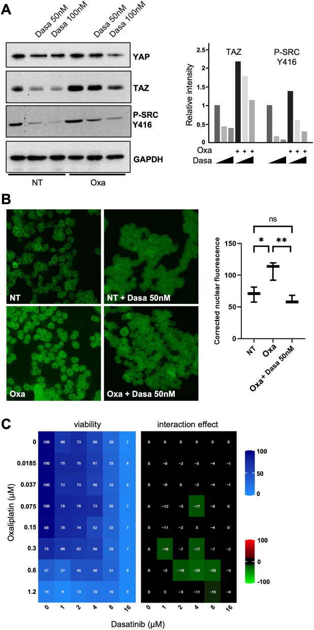
Fig. 6.
Src inhibition by Dasatinib reduces HCT116 cells sensitivity to oxaliplatin. A Western blot analysis showing protein expression of YAP and TAZ in HCT116 treated, or not, with oxaliplatin (0.5 µM) and/or Dasatinib (50 nM and 100 nM). Phopsho-SRC blotting was used to evaluate the inhibition of SRC activity using Dasatinib. GAPDH was used as a loading control (n = 3). Quantification of the blots (performed using Image J software) is shown on the right side of the figure. B Immunofluorescence experiments performed in HCT116 cells treated, or not, with oxaliplatin (0.5 µM) and/or Dasatinib (50 nM) monitoring YAP (top panels) and TAZ (bottom panels) nuclear localization (red). DAPI (blue) was used to stain DNA and the nuclei. Quantification of both stainings are shown on the right side of the figures and are represented as the corrected nuclear fluorescence. Data are represented as the mean ± SEM (n = 3). Unpaired two-tailed Student’s t-test; ****p < 0.0001. C HCT116 colorectal cancer cell lines were incubated with increasing concentrations of oxaliplatin and Dasatinib. Cell viability was assessed with the SRB assay in 2D to obtain the viability matrix. Drug concentrations were as follows: Dasatinib (from 1 to 16 µM) and oxaliplatin (from 0.0185 to 1.2 µM). The synergy matrix was calculated as described in “Materials and methods” section

