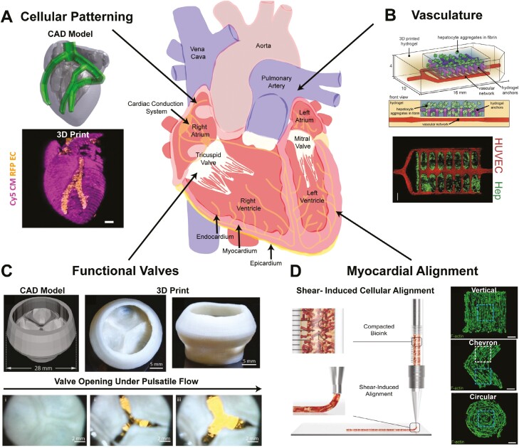
Figure 1.
Capabilities of bioprinting to build different features of the heart. Bioprinting has enabled the (A) spatial patterning of cells like endothelial cells (red) within a cardiomyocyte structure (pink).27 (B) Vasculature has been created using light-based bioprinting and subsequently endothelialized (red), forming perfusable channels within hepatocyte aggregates (green).28 (C) Creation of functional acellular components has been accomplished, including trileaflet heart valves that open and close under physiologically relevant flows and pressures.5 (D) Reconstruction of microscale structural features, like myocardial alignment, has been accomplished utilizing shear stress through the needle during extrusion-based bioprinting.29

