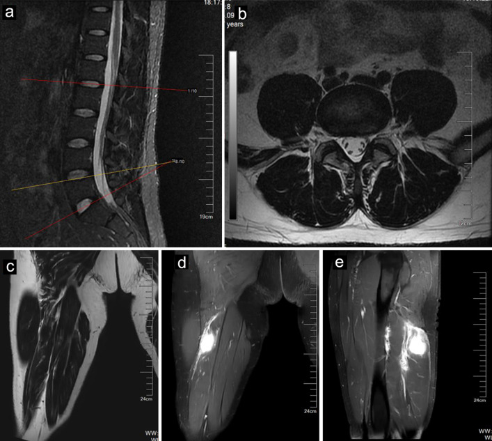
Figure 1.
MR images of a 40-year-old male with IMT. Sagittal (a) and axial (b) MR images of the lumbar spine showing a mildly herniated disc at the L4-L5 level. The tumor (arrows) showed mild hyperintensity on T1-weighted imaging (c) and hyperintensity on fat-suppressed T2-weighted imaging (d). The tumor (arrows) showed enhancement after contrast agent injection (e). IMT: inflammatory myofibroblastic tumor; MR: magnetic resonance.

