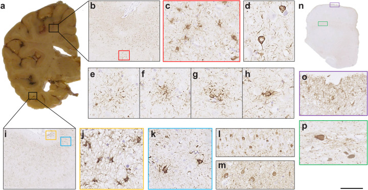
Extended Data Figure 5. R406W mutation in MAPT: Immunohistochemical localisation of tau inclusions in case 2.
a, Mild atrophy of the frontal cortex, severe atrophy of the temporal cortex and underlying white matter, with marked reduction in bulk of the hippocampus. Anterior and temporal horns of the lateral ventricle are dilated; b, tau pathology in the anterior frontal cortex; c, some stained cells resemble tufted or thorn-shaped astrocytes; d, abundant neuronal inclusions and neuropil threads; e, astrocytic plaque; f,g, structures in-between astrocytic plaques and tufted astrocytes; h, tufted astrocyte; i,k, Tau pathology in the lateral temporal cortex was similar to that in the anterior frontal cortex; l, CA4 region of the hippocampus; m, dentate gyrus; j, subpial astrocytic tau pathology at the depth of a sulcus in the lateral temporal cortex. n, Low-power view of the midbrain; o, subpial astrocytic tau pathology; p, neuronal tau staining in the substantia nigra. AT8 antibody. Scale bar: b, 750 μm; c,k, 70 μm; d, 40 μm; e,f,g,h, 30 μm; i, 670 μm; j, 50 μm; l. 100 μm; m, 110 μm; n, 5.5 mm; o,p, 90 μm.

