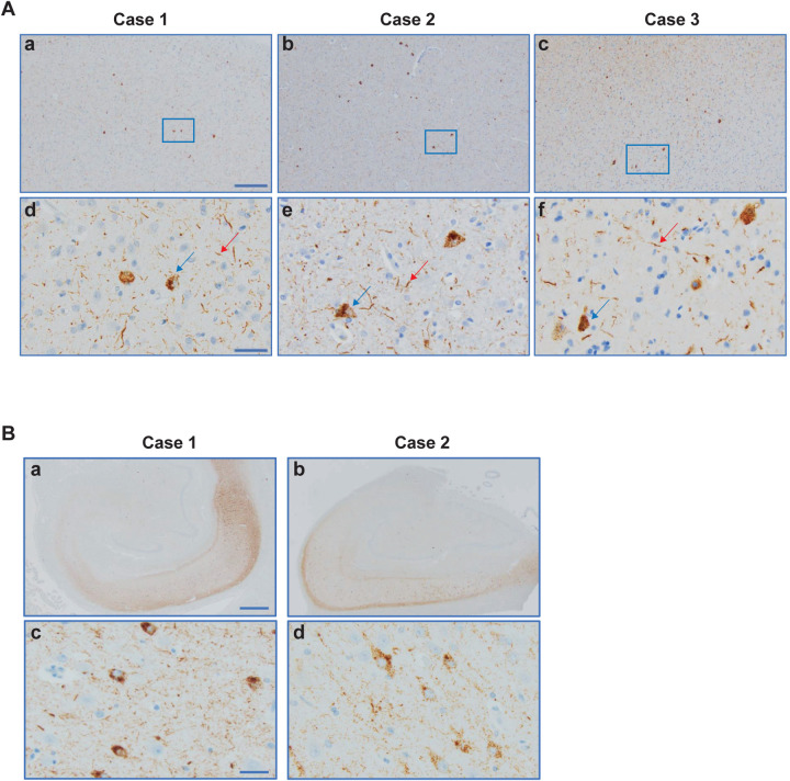
Extended Data Figure 1. V337M mutation in MAPT: Immunohistochemical localisation of tau inclusions in frontal cortex and hippocampus.
A. (a-f) Tau pathology in grey matter. Higher magnifications of tissue areas within the insets in a-c are shown in d-f. Intraneuronal inclusions (blue arrows) and neuropil threads (red arrows) are in evidence. Antibody: AT8. Scale bars: 200 μm (a-c); 40 μm (d-f).
B. a,b, Low-power view; c,d, neurofibrillary tangles and neuropil threads in the pyramidal cell layer. Antibody: AT8. Scale bars: 1,000 μm (a,b) and 40 μm (c,d).

