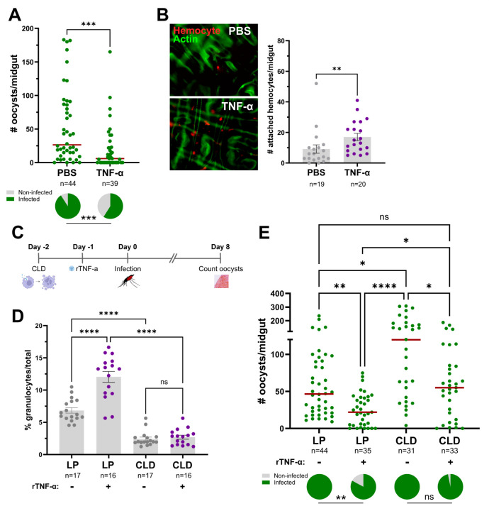
Figure 5. TNF-mediated parasite killing targets ookinete invasion and is mediated in part by granulocyte function.
(A) Adult female mosquitoes were injected with 1XPBS (control) or 50ng of rTNF-α prior to infection with P. berghei. Oocyst numbers and infection prevalence were evaluated at 2 days post infection to measure early oocyst numbers and the success of ookinete invasion. (B) Immunofluorescence images of DiI-stained hemocytes (red) attached to the mosquito midgut (counterstained with phalloidin, green) approximately 24 hrs post-treatment with 1xPBS (control) or 50ng of rTNF-α. The number of attached hemocytes was quantified for each respective treatment with the dots corresponding to the number of hemocytes attached to each individual midgut examined. To address the role of granulocytes in TNF-mediated parasite killing, mosquitoes were first injected with either clodronate liposomes (CLD) to deplete granulocytes or control liposomes (LP), then 24 hours later surviving mosquitoes were treated with 50ng rTNF-α or 1X-PBS (C). Each group was then challenged with P. berghei and oocyst numbers were examined on Day 8 post-infection. (D) Before infection, the effects of clodronate treatment on the percentage of granulocytes was examined in the presence or absence of rTNF-α to confirm our experimental approach. (E) Infection outcomes following granulocyte depletion and rTNF-α treatment, with oocyst numbers and infection prevalence evaluated at 8 days post infection. For both D and E, “+” denotes treatment with rTNF-α, while “-“ indicates treatment with 1X-PBS. For all experiments, the dots represent the respective measurements from an individual mosquito. The red horizontal lines represent the median oocysts numbers, while infection prevalence (% infected/total) is depicted as chart pies below each figure containing infection data. Data were combined from three or more independent experiments. Statistical significance was determined using either Mann-Whitney (individual comparisons) or Kruskal-Wallis with a Dunn’s multiple comparison test (multiple comparisons) to assess oocyst numbers, the number of attached hemocytes, or the percentage of granulocytes. A Fisher’s Exact test was performed to measure differences in infection prevalence. Asterisks denote statistical significance (* P < 0.05, ** P < 0.01, *** P < 0.001, **** P < 0.0001). ns, not significant; n= numbers of individual mosquitoes examined.

