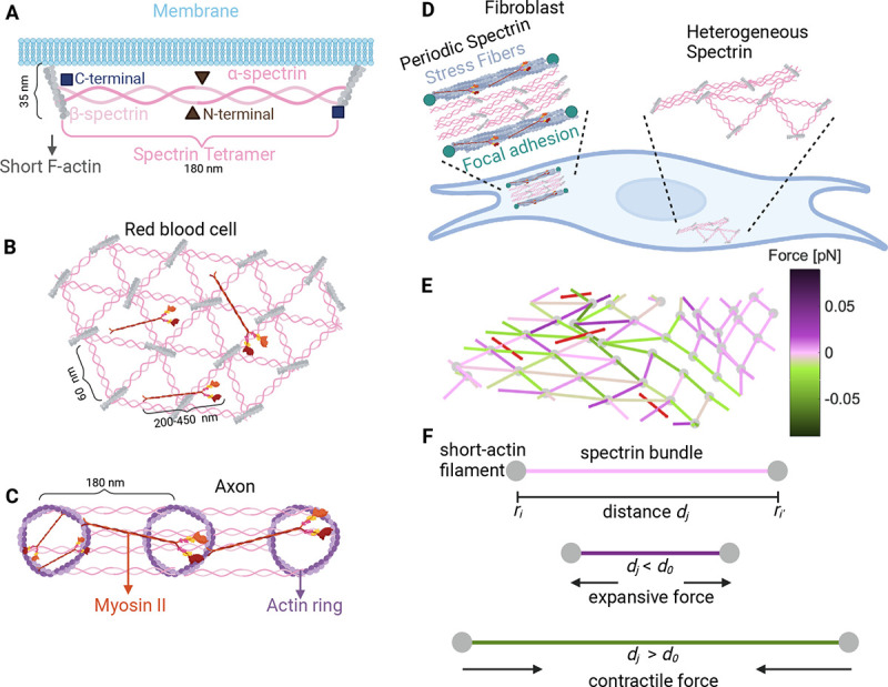
Figure 1: Different configurations of the membrane skeleton.

A) A spectrin tetramer spanning between short actin filaments. B) The hexagonal actin-spectrin meshwork configuration in red blood cells. Myosin generates contractility that may preserve the cell shape (18). C) Periodic actin-spectrin meshwork configuration in axons. Myosin heavy chains crosslink adjacent actin rings, likely providing tension. Myosin may also span individual rings providing contraction (15). D) In fibroblasts, the actin-spectrin meshwork has a heterogeneous and dynamic configuration (11). E) Schematic of the simulated 3D network model. The red lines correspond to myosin, grey nodes to short F-actin, and edges to spectrin, color-coded for the force generated by the spring element. F) Schematic representation of the forces generated by the spectrin edges when their length differs from the resting length.
