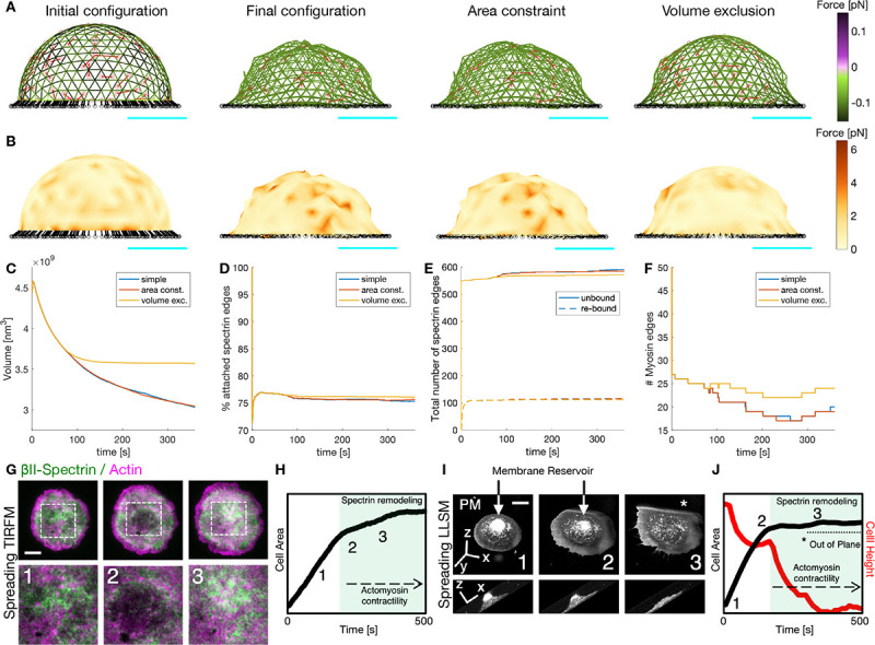
Figure 8: Actin-spectrin meshwork dynamics on an adhered cell.

A) Initial configuration of the actin-spectrin spherical meshwork and 360 seconds of the simulation, with area constraint and volume exclusion. Edges are color-coded for the force generated by the spring potential energy of the spectrin edges. Red lines correspond to myosin edges. The black lines represent connecting edges and black circles, connecting nodes. Cyan line is a scale bar corresponding to 1 μm. B) Meshwork in A but color-coded for the force generated by the membrane. Here, . Time evolution of the volume (C), percentage of attached spectrin edges (D), total number of spectrin edges unbound and re-bound (E), and number of myosin edges (F). G) Cell spreading analysis at the cell body (zooms corresponding to the dashed white boxes), displayed by live TIRFM images (green: GFP-βII-spectrin, magenta: RFP-actin, scale bar: 10 μm). Relevant events observed between independent experiments are shown (1–3), in particular, endogenous actin node formation and correspondent βII-spectrin behavior. H) Projected Cell Area analysis over time and the relative positioning of frames 1–3 presented in G are shown in the graph. Activation of actomyosin contractility and spectrin remodeling during the slow-growth phase of spreading (P2) is highlighted in green. Figures adapted from (10). I) Cell spreading imaged by Lattice Light Sheet Microscopy in MEF transfected with the membrane reporter Scarlet-PM(Lck), scale bar: 10 μm. Relevant frames 1–3 are reported in the orthogonal view (whole cell) and in the lateral projection (to highlight cell height). The membrane reservoir is present on the top of the cell body and dissolved during the slow-growth phase of spreading (P2). J) Projected Cell Area (black) and Cell height (red) analysis over time and the relative positioning of frames 1–3 presented in C are shown in the graph. Activation of actomyosin contractility and spectrin remodeling during the slow-growth phase of spreading (P2) is highlighted in green, correlating to the flattening of the cell body. The portion of the cell that is excluded from the illumination plane is indicated by the asterisk (*).
