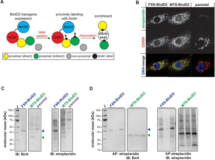
Figure 1. Mitochondrial localization and biotinyl ligase activity of BioID2 transgenes.
(A) BioID2 was cloned in-frame with carboxy terminus of human FXN, Prdx3, and the MTS of COX8a for detection and enrichment of proximal interacting proteins. (B) Immunofluorescent labeling of parental and stably transfected A549 cells expressing BioID2 transgenes that were pulsed with 50 μM biotin for streptavidin (green) and COXIV (red) to visualize transgene expression, subcellular localization, and biotinyl ligase activity. Dapi was used as a nuclear counterstain (blue in DNA-merge). SDS-PAGE/immunoblots of (C) whole cell lysates or (D) streptavidin affinity purified A549 BioID2 cell lysates with anti-BirA antibody or streptavidin for transgene detection and protein biotinylation. Colored arrowheads indicate detection of predicted-sized transgenes.

