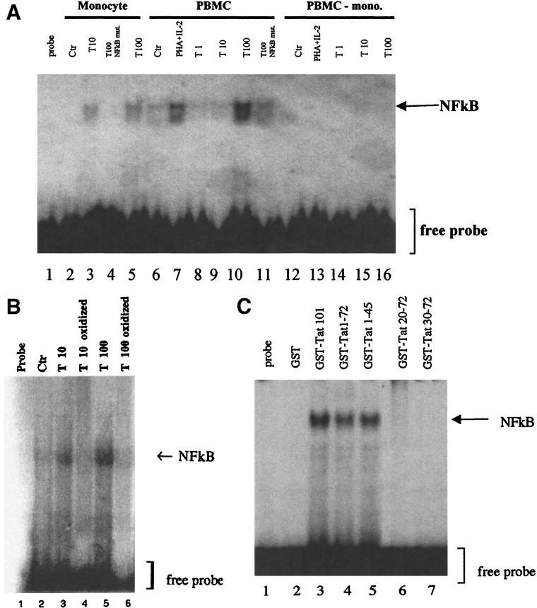
FIG. 7.
Activation of NF-κB by Tat in human monocytes. (A) Activation of NF-κB determined by EMSA. Nuclear protein extracts of monocytes, total PBMC, or monocyte-depleted PBMC (PBMC-mono.) were treated with Tat at 1 nM (lanes 8 and 14), 10 nM (lanes 3, 9, and 15), or 100 nM (lanes 5, 10, and 16) or with PHA (3 mg/ml) plus IL-2 (10 U/ml) (lanes 7 and 13) for 16 h and then incubated with a sequence containing the wild-type NF-κB site. The specificity of interaction was tested by using a 32P-labeled mutated NF-κB probe (NF-κB mut.) incubated with extracts from cells treated with 100 nM Tat (lanes 4 and 11). These results are representative of two independent experiments done on cells from two donors. Ctr, control. (B) Specificity of Tat-mediated NF-κB activation. Nuclear protein extracts of monocytes treated with native Tat (10 and 100 nM) (lanes 3 and 5, respectively) or with oxidized Tat (10 and 100 nM) (lanes 4 and 6, respectively) for 16 h were incubated with the 32P-labeled wild-type NF-κB probe as described above. (C) Localization of the domain of Tat involved in NF-κB activation. Nuclear protein extracts of monocytes treated with wild-type GST-Tat 1-101 (lane 3) or Tat-deleted mutants (GST-Tat 1-72, GST-Tat 1-45, GST-Tat 20-72, and GST-Tat 30-72) at 10 nM (lanes 4 to 7) or as a negative control with GST (10 nM) for 16 h were incubated with 32P-labeled NF-κB probe.

