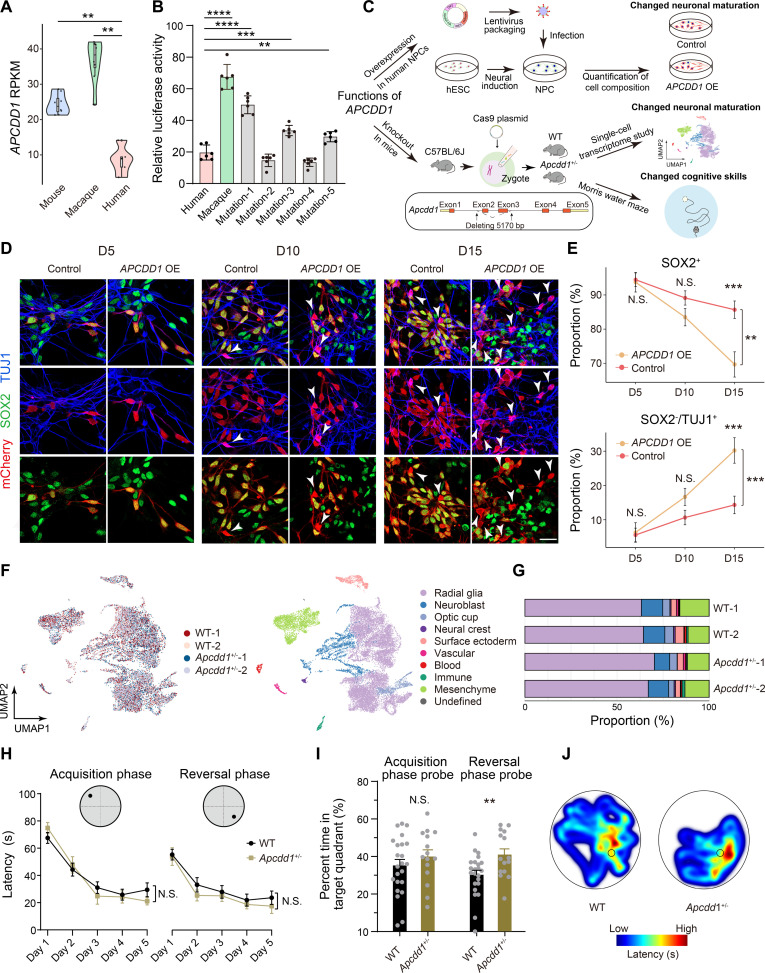
Fig. 7. A human-specific inversion contributes to human uniqueness in brain development.
(A) Violin plots showing the expressions of APCDD1 in the brains of humans, macaques, and mice at the mid- to late-fetal stages. Wilcoxon rank-sum tests, **P < 0.01. (B) Relative luciferase activities of human APCDD1 promoter (Human), macaque APCDD1 promoter (Macaque), and human APCDD1 promoter with mutations (Mutation-1 to Mutation-5). Student’s t test, **P < 0.01, ***P < 0.001, ****P < 0.0001. (C) The design of experiments for APCDD1 functions. (D) Representative immunostaining of SOX2 (progenitors), TUJ1 (postmitotic neurons), and mCherry (virus-infected cells) in the assays of wild-type NPCs (Control) and NPCs with APCDD1 overexpression (APCDD1 OE), at different protocol days (D5, D10, and D15) after lentiviral infection. White arrowheads, SOX2−/TUJ1+ cells. Scale bars, 20 μm. (E) Proportions of SOX2+ progenitors and SOX2−/TUJ1+ neurons in Control and APCDD1 OE at different protocol days (D5, D10, and D15). Two-way ANOVA, **P < 0.01, ***P < 0.001. (F) UMAP plots of scRNA-seq from brains of wild-type (WT-1 and WT-2) and Apcdd1+/− mice (Apcdd1+/−-1 and Apcdd1+/−-2) at E10.5, grouped by genotypes (left) or cell types (right). (G) Proportion of each cell type in brains of WT-1, WT-2, Apcdd1+/−-1, and Apcdd1+/−-2. (H) The learning curves of the wild-type (WT, n = 23) and Apcdd1+/− (n = 15) mice in the acquisition and reversal phases. Two-way ANOVA. N.S., not significant. (I) Time spent in the target quadrant by WT and Apcdd1+/− mice in the probe trials after the acquisition and reversal phases. Two-sided Student’s t test, **P < 0.01, N.S., not significant. (J) Representative plots of escape latency for WT and Apcdd1+/− mice in the probe trials after the reversal phase of learning. Data are represented as the means ± SEMs.

