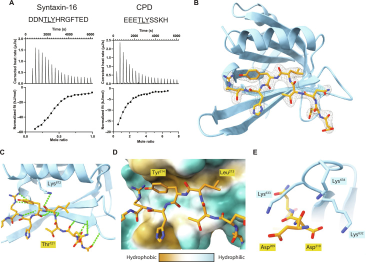
Fig. 4. Structure of TBC1D23 C-terminal domain bound to a syntaxin-16 peptide.
(A) ITC trace and fitted curve of the indicated peptides from syntaxin-16 (left) and CPD (right) binding to TBC1D23 C-terminal domain. Source data are in data S2. (B) Structure of the TBC123 C-terminal domain (sky blue) and syntaxin-16 (209 to 217) (gold) with the corresponding electron density map of the latter. (C) Close-up of the hydrogen bonding network between syntaxin-16 (gold) and TBC1D23 (sky blue). Lys672 hydrogen bonds to the backbone of syntaxin-16. Hydrogen bonds are in green. (D) Hydrophobicity surface plot of TBC1D23 showing the packing of residues of syntaxin-16 (gold) into two hydrophobic pockets. (E) Close-up of Asp209 and Asp210 in syntaxin-16 (gold) interacting with Lys632 to Lys634 in TBC1D23 (sky blue).

