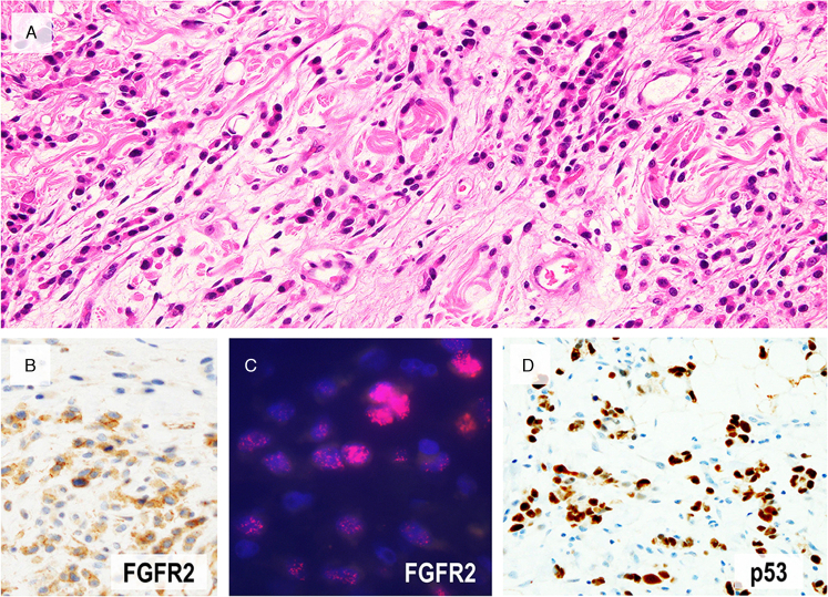
FIGURE 4.
A case with FGFR2 amplification. (A) A representative image of invasive area showing diffuse-type histology. In this advanced tumor with FGFR2 amplification, overexpression by immunohistochemistry (B) and amplification by FISH (C, red signals) was confirmed in diffuse-type components but not in very well-differentiated component. This tumor showed diffuse positivity for p53 (D).

