Figure 2.
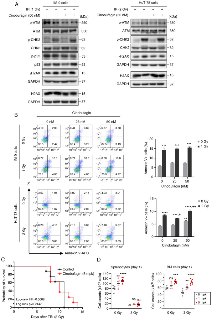
Radioprotective effects of cinobufagin. (A) IM-9 and HuT 78 cells were treated with or without 50 nM of cinobufagin for 1 h, then irradiated with the indicated dose of radiation and incubated for 24 h. The expression levels of ATM, CHK2, p53 and H2AX phosphorylation were evaluated using western blotting. (B) IM-9 and HuT 78 cells were treated with or without cinobufagin (25 or 50 nM) for 1 h prior to irradiation with the indicated radiation doses for 24 h. The cells were analyzed for Annexin V/PI staining using flow cytometry. Representative bar graphs show the mean ± SEM of three independent experiments; **P<0.01 and ***P<0.001 compared with the control, +P<0.05 and ++P<0.01 compared with the irradiated control. (C) Cinobufagin or the vehicle (1.1% DMSO in PBS) were administrated intraperitoneally 24 h prior to total body radiation (8 Gy) on C57BL/6 mice (n=8 mice/group) and survival was observed for 15 days after exposure. (D) Cinobufagin or the vehicle were administrated intraperitoneally 24 h prior to total body radiation (3 Gy) on C57BL/6 mice (n=5 mice/group). The spleen (left panel) and BM (right panel) were harvested at 24 h following radiation, and the number of splenocytes and BM cells were counted by Trypan blue exclusion. Data shown are the mean ± SEM. Statistical significance was represented for each control group (**P<0.01, ***P<0.001, ****P<0.0001). ns, not significat; IR, ionizing radiation; TBI, total body irradiation; ATM, ataxia telangiectasia mutated; CHK2, checkpoint kinase 2; H2AX, H2A histone family member X.

