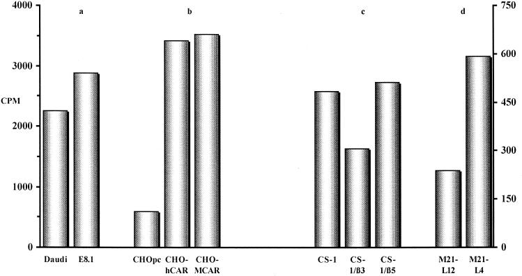
FIG. 5.
CAV-2 attachment to CAR, MHC-1 α2 domain, and αvβ5 integrins. Daudi (a), CHO (b), CS-1 (c), and M21-L (d) cells and their derivatives were incubated with 35S-CAV in order to determine the binding capacities relative to the parental cell line. The data are presented as the total counts per minute per cell type. The background counts (cells blocked with cold CAV-2 prior to incubation with 35S-CAV) were ca. 100 cpm for CHO- and CS-1-derived cells and 250 and 25 cpm for Daudi and E8.1 cells, respectively. For Daudi, E8.1, and the CHO derivatives, the y axis is on the left (0 to 4,000 cpm). For CS-1 and M21-L cells, the y axis is on the right (0 to 600 cpm). The data are the means of two samples, with an error of <10% for each sample.

