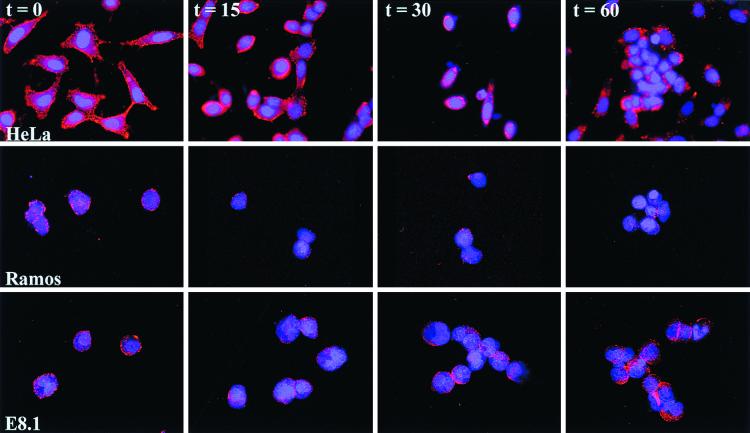
FIG. 8.
CAV-2 attachment and entry. CAV-Cy3 was incubated with Ramos, E8.1, and HeLa cells in order to identify the point of inhibition for CAV-2 transduction. Cells were fixed at the indicated times. CAV-Cy3 appears as red points, and the cell nuclei were counterstained with DAPI (4′,6′-diamidino-2-phenylindole).

