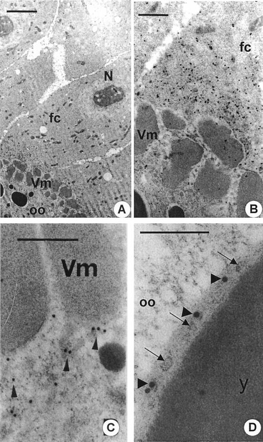
FIG. 7.
Immunocytochemical detection of Gag viral antigens. (A) The follicle cell-oocyte border from a stage 9 RevI ovarian follicle tested with anti-Gag antibody. fc, posterior follicle cell; N, follicle cell nucleus; oo, oocyte; Vm, vitelline membrane. Bar, 4 μm. (B) Enlargement of panel A to show numerous 20-nm gold grains of the secondary antibody along the apical end of the follicle cell. Bar, 1 μm. (C) Portion of the cortical ooplasm from a stage 10 RevI ovarian follicle showing gold grains (arrowheads) due to anti-Gag antibody along the oolemma. Bar, 0.5 μm. (D) A forming yolk granule (y) from a stage 9 RevI ovarian follicle. Note the presence of gold grains due to anti-Gag antibody (arrowheads) over the superficial layer among viral particles (arrows). Bar, 0.4 μm.

