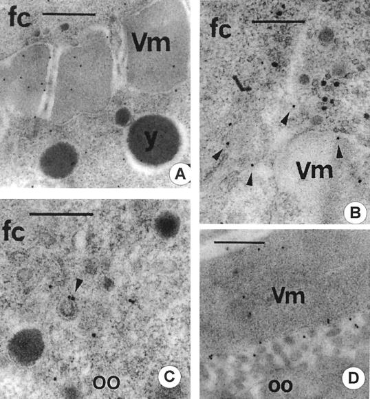
FIG. 8.
Immunocytochemical detection of Env viral antigens. (A) The follicle cell (fc)-oocyte border from a stage 10 RevI ovarian follicle exposed to anti-Env antibody. Gold grains are dispersed over the vitelline membrane (Vm). y, yolk granule. Bar, 0.5 μm. (B) The apical end of a posterior follicle cell from a stage 9 RevI ovarian follicle showing several gold grains (arrowheads) along the plasma membrane. Bar, 0.4 μm. (C) The posterior-most cortical ooplasm from a stage 9 RevI ovarian follicle tested with anti-Env antibody. Arrowhead, gold-labeled coated vesicle. oo, oocyte. Bar, 0.4 μm. (D) Portion of a stage 11 RevI ovarian follicle showing the vitelline membrane and the underneath oolemma. Vitellogenic uptake has ceased by this developmental stage in D. melanogaster, and yet gold grains due to the anti-Env antibody are still seen bound along the microvilli of the oolemma. Bar, 0.6 μm.

