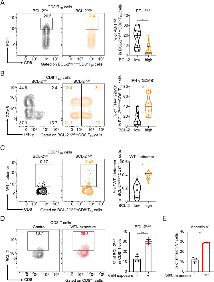
Fig. 2. BCL-2high CD8+ TEM cells have a progenitor/non-exhausted, cytotoxic, and tumor-specific phenotype, and VEN exposure enhances the anti-tumor immune response of CD8+ T cells.
A–C Proportion of PD-1high (n = 11) (A), IFN-γ+GZMB+ (n = 8) (B), and WT-1 tetramer+ (n = 3) (C) cells in BCL-2high/low CD8+ TEM cells. Samples were obtained from post-VEN therapy patients, and flow cytometry analysis was performed on PBMCs for PD-1 and WT-1 data, and on bone marrow samples for cytokine data. D Proportion of BCL-2high cells in CD8+ T cells. Healthy donor PBMCs stimulated with anti-CD3 monoclonal antibody and IL-2 (200 IU/ml) were cultured in medium containing 0.1 µM VEN for 96 hours and then analyzed with flow cytometry (n = 3). E In vitro killing assay. CellTrace Yellow-labeled KG-1 cells were cocultured with VEN-exposed T cells. Twenty-four hours later, cells were stained with Annexin V and analyzed with flow cytometry (n = 3). Unpaired t-tests were used for statistical calculations.; *P < 0.05; **P < 0.01.

