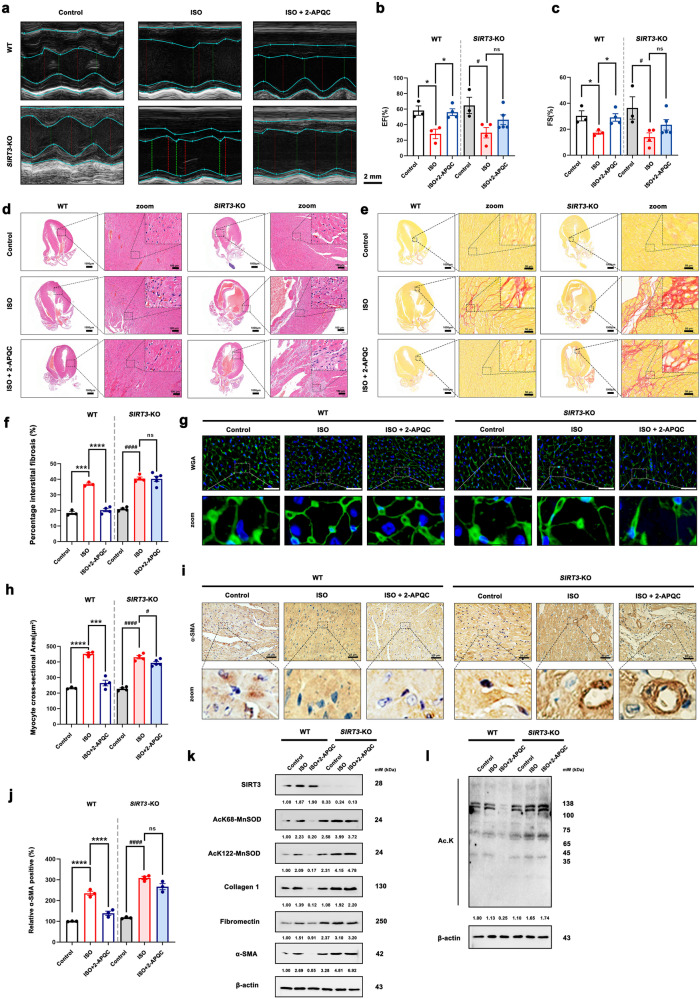
Fig. 4.
2-APQC could not alleviate ISO-induced HF in SIRT3 knockout mouse model. a–c Representative image of echocardiogram of indicated groups. n = 3. d Represents the result of HE staining in heart tissues, scale bar = 1 mm; scale bar = 100 μm. e, f Sirius Red staining in heart tissues. Data are present as mean ± s.e.m, n = 3. g, h wheat germ agglutinin (WGA) staining in heart tissues, scale bar = 20 μm. Data are present as mean ± s.e.m, n = 3. i, j Represents the result of α-SMA staining in heart tissues, scale bar = 25 μm. Data are present as mean ± s.e.m, n = 3. k, l Detection and quantification of the expression of SIRT3, ac-MnSOD2 (K68), ac-MnSOD2 (K122) fibronectin, collagen 1 and α-SMA by Western blot. β-actin, loading control. ns, no significance, *p < 0.05, **p < 0.01, ***p < 0.001, ****p < 0.0001; #p < 0.05, ##p < 0.01, ###p < 0.001, ####p < 0.0001. Nico nicotinamide, ISO isoproterenol, Hon Honokiol, Met metoprolol

