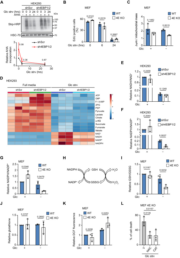
Fig. 2. 4EBP1/2 maintain antioxidant power and preserve the redox balance under glucose starvation.
A Control (shScr) and sh4EBP1/2 HEK293 cells were grown in complete medium or glucose starved (Glc strv) for the indicated times and labeled with azidohomoalanine (AHA). Levels of AHA-labelled proteins were detected by immunoblotting. B WT and 4E KO MEF were grown in complete medium or glucose starved (Glc strv) for the indicated times, labeled with EdU and analyzed by flow cytometry. C WT and 4E KO MEF were grown in complete medium or glucose (Glc) starved for 24 h. Mitochondrial membrane potential (Δψm) with TMRE staining and mitochondrial mass were measured. D Control (shScr) and sh4EBP1/2 HEK293 cells were grown in complete medium or glucose (Glc) starved for 24 h and the corresponding metabolites were measured by LC-MS. E NAD+ and NADH levels measured in (C) were plotted as NADH/NAD+ ratio. F NADP+ and NADPH levels measured in (C) were plotted as NADPH/NADP+ ratio. G WT and 4E KO MEF were grown in complete medium or glucose (Glc) starved for 24 h, and NADP+ and NADPH levels were measured. H Scheme of the usage of NADPH in recycling oxidized glutathione for H2O2 detoxification. (I) WT and 4E KO MEF were grown as in (C), and reduced and total glutathione were measured and expressed as the ratio of reduced (GSH) to oxidized (GSSG) glutathione. J WT and 4E KO MEF were grown as in (C), and total glutathione was measured. K WT and 4E KO MEF grown as in (C) were labelled with CM-DCFDA and analyzed by flow cytometry. L 4E KO MEF were grown in glucose starved medium (Glc strv) and treated with vehicle (V), N-acetyl cysteine (NAC) or Catalase (CAT) for 48 h. Cell death was measured by PI staining and flow cytometry. Data are shown as the mean ± SD. Statistics: unpaired one-sided Student’s t test (A–C, E–G, I–L); n = 3 independent experiments for (A–C, E–G, I–L). Source data are provided as a Source Data file.

